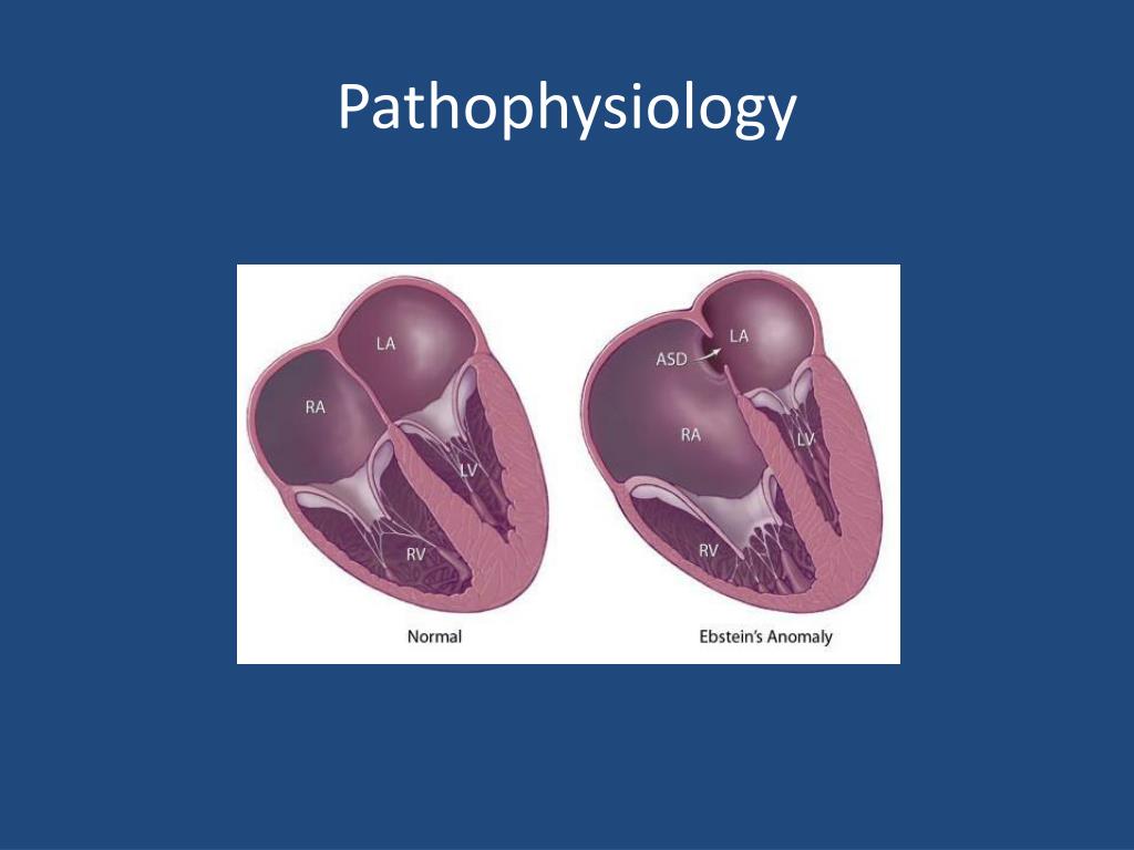
Morphology of the right side of a normal heart.

In addition, the tension apparatus of the septal leaflet is attached to the coarsely trabeculated septal surface, and the pulmonary valve is supported by a complete muscular sleeve separating it from the tricuspid valve. The tricuspid valve consists of three leaflets designated anterior-superior, inferior, and septal, and its hinge point is located at the annulus. Ī normal morphologic right ventricle ( Figure 1a and b) is divided into inlet, apical trabecular, and outlet components and has a tricuspid valve with its orifice pointing toward the ventricular apex. Furthermore, an Ebstein’s anomaly may be associated with an atrial septal defect, pulmonary atresia, or congenitally corrected transposition of the great arteries (ccTGA) and less commonly with pulmonary stenosis, a ventricular septal defect, or an atrioventricular septal defect. In addition, Ebstein’s malformation is often accompanied by valvar dysplasia, anomalies of the tension apparatus, and myocardial anomalies, and in severe cases, the dilated thin-walled, atrialized inlet component is divided from the apical tubercular and outlet components by a muscular shelf. The chief distinguishing feature of an Ebstein’s malformation is the positioning of the hinge point of the tricuspid valve into the right ventricular cavity with an apical and anterior rotated appearance toward the right ventricular outflow tract rather than at its normal location at the atrioventricular junction or annulus, and this is due to failure of the septal and inferior leaflets to delaminate from the underlying myocardium. Ģ.1 Ebstein’s anomaly is an anomaly with both myocardial and valvular defects Presentation of this malformation varies widely from neonates in extremes to an incidental finding during physical examination in an otherwise asymptomatic adult secondary to the anatomic severity of the tricuspid valve and the associated heart malformations. This malformation is thought to be due to the defects in the process of “delamination from the underlying myocardium” during the development of the tricuspid valve. Ebstein’s anomaly is more than an issue with inferior displacement and rotation of the tricuspid valve, as this anomaly may also involve abnormalities of the right ventricular myocardium and in some cases the left ventricle. The patient, Joseph Prescher, was a 19-year-old worker who presented with cyanosis, dyspnea, palpitations, cardiomegaly, and distended jugular veins. Ebstein’s anomaly was first described by Wilhelm Ebstein with the autopsy findings of abnormal tricuspid valves in 1866. Given the low incidence of this defect, some centers will have limited experience managing patients with Ebstein’s anomaly, suggesting the potential importance of regionalizing care in patients with more complex physiology. In a report from the Society of Thoracic Surgery (STS) Congenital Heart Surgery Database from 2010 to 2016, there were 494 patients with Ebstein’s anomaly who received index operations in 95 centers. This tricuspid valve repair is effective and durable for the majority of patients.Įbstein’s anomaly of the tricuspid valve is a rare congenital heart malformation that accounts for about 0.5% of all congenital heart defects and 0.005% of all live births. The cone procedure for reconstruction for Ebstein’s anomaly can be performed with low mortality and morbidity.
#Ebstein anomaly tv
In this chapter, we review the surgical maneuvers that we have used to obtain the best functional TV in cases with several anatomic variations of Ebstein’s anomaly. The result mimics normal TV anatomy, which is an improvement compared to previously described procedures that result in a monocusp valve coaptation with the ventricular septum. This operation aims to undo most of the anatomic TV defects that occurred during embryologic development and to create a cone-like structure from all available leaflet tissue. developed a surgical technique involving cone reconstruction of the TV. The wide variety of anatomic and pathophysiologic presentations of Ebstein’s anomaly has made it difficult to achieve uniform results with surgical repair, resulting in the development of many different surgical techniques for its repair.

Ebstein’s anomaly of the tricuspid valve is a cardiac malformation characterized by downward displacement of the septal and inferior tricuspid valve (TV) leaflets, redundant anterior leaflets with a sail-like morphology, dilation of the true right atrioventricular annulus, TV regurgitation, and dilation of the right atrium and ventricle.


 0 kommentar(er)
